
The dominant medical model frames demyelination as a structural pathology, primarily managed through pharmacological interventions. However, emerging evidence from quantum biology and mitochondrial biophysics presents a radically different view: demyelination is a consequence of quantum signal degradation, mitochondrial dysfunction, and circadian incoherence. This unified theory integrates photonic signaling, redox biology, and quantum coherence to explain, and potentially reverse, neurological decline at the first-principles level.
The Myelin–Mitochondria–Microtubule Axis
Myelin sheath integrity depends on high-fidelity ATP production, structured water networks, and ultraweak photon emissions (UPEs) generated by mitochondria. These photons act as biophysical signals to guide:
-
Oligodendrocyte precursor cell (OPC) mitosis
-
Microtubule self-assembly
-
Synaptic pruning
-
Neural regeneration
Mitochondria are quantum semiconductors. They rely on a tightly controlled redox environment to emit UPEs in the UV-visible spectrum. When light exposure, redox potential, and water chemistry are optimized, mitochondria produce coherent UPEs that regulate the neural cytoskeleton and support consciousness (Van Wijk & Van Wijk, 2005).
Diseases like Multiple Sclerosis (MS), Alzheimer's, and Parkinson’s show clear patterns of mitochondrial dysfunction, reduced ATP production, elevated ROS, and disrupted circadian rhythm. In MS, demyelination occurs after microglial activation and inflammatory cytokine release, but upstream from this is mitochondrial redox collapse and structural disintegration of OPC support systems (Campbell et al., 2014).
The Welding Analogy: Myelin as a Light-Forged Circuit
Arc welding requires:
-
Precision in voltage/amperage (like mitochondrial PMF)
-
Clean substrates (akin to mtDNA and redox purity)
-
Proper shielding (analogous to antioxidant buffering and deuterium-depleted water)
-
Light feedback (UPEs from cytochrome c oxidase)
Just as contaminated electrodes produce brittle welds, environmental pollutants, heavy metals, tattoos, non-native EMFs, and dopants like nanoparticles disrupt mitochondrial redox balance and UPE coherence leading to defective myelin. Tattoos, for example, introduce carbon-based dopants that increase static charge and oxidative stress, especially when exposed to high-frequency EMFs. This disrupts ATPase function and impairs OPC differentiation (Kruse, 2023).
Alzheimer’s patients demonstrate impaired mitochondrial membrane potential and faulty mitophagy. The photonic feedback required for neuronal microtubule maintenance is degraded, leading to tau hyperphosphorylation and cytoskeletal collapse (Swerdlow et al., 2014).
DDW, UPEs, and the Quantum Gatekeeper
Deuterium-depleted water (DDW) optimizes proton tunneling through ATP synthase, stabilizes hydrogen bonding networks, and reduces oxidative stress. This enhances the purity and coherence of mitochondrial UPEs.
Vitamin D, synthesized from sunlight not oral supplements activates the vitamin D receptor (VDR) located on the inner mitochondrial membrane. This fine-tunes the urea cycle, regulates calcium influx, and supports TCA intermediates essential for lipid synthesis and myelination. Oral supplements bypass this light-triggered circadian interface, creating signaling mismatches.
Loss of UV light exposure leads to VDR uncoupling, reduced melatonin and dopamine synthesis, and dysfunctional OPCs. This explains why demyelination risk is higher in populations with indoor lifestyles and lower sun exposure (Pierrot-Deseilligny & Souberbielle, 2017).
Shannon’s information theory applies here: fewer coherent light inputs mean higher noise-to-signal ratios in cellular decision-making. Mitochondria lose the ability to discriminate between growth and apoptosis pathways, increasing heteroplasmy and neural cell death.
Consciousness: From UPEs to Qualia
Biophotons produced by mitochondrial respiration interface with cerebrospinal fluid (CSF), structured water, and neural microtubules. These UPEs act as wireless quantum instructions for:
-
Cell division (via ubiquitin-coupled signaling)
-
Cytoskeletal coherence
-
Wakefulness and attention
In Alzheimer’s and ALS, this coherence breaks down before memory loss. Myelin deteriorates, tau proteins aggregate, and axonal transport fails not from genetic causes alone but from chronically insufficient photonic input and redox instability (Mattson & Liu, 2002).
Neuromelanin, found in high concentrations in the substantia nigra, absorbs UV and visible light. Its breakdown in Parkinson’s correlates with the progressive loss of dopamine neurons, suggesting that demyelination and neurodegeneration may result from failing light-capture machinery in the brain (Zecca et al., 2004).
Clinical Application: From Light to Repair
Sunlight is the master regulator:
-
AM UV entrains circadian clocks, builds mitochondrial charge, and triggers vitamin D synthesis
-
PM infrared stabilizes structured water, enhances ATP production, and repairs DNA
-
Full-spectrum daily exposure resets melanin, redox, and VDR activation across all tissues
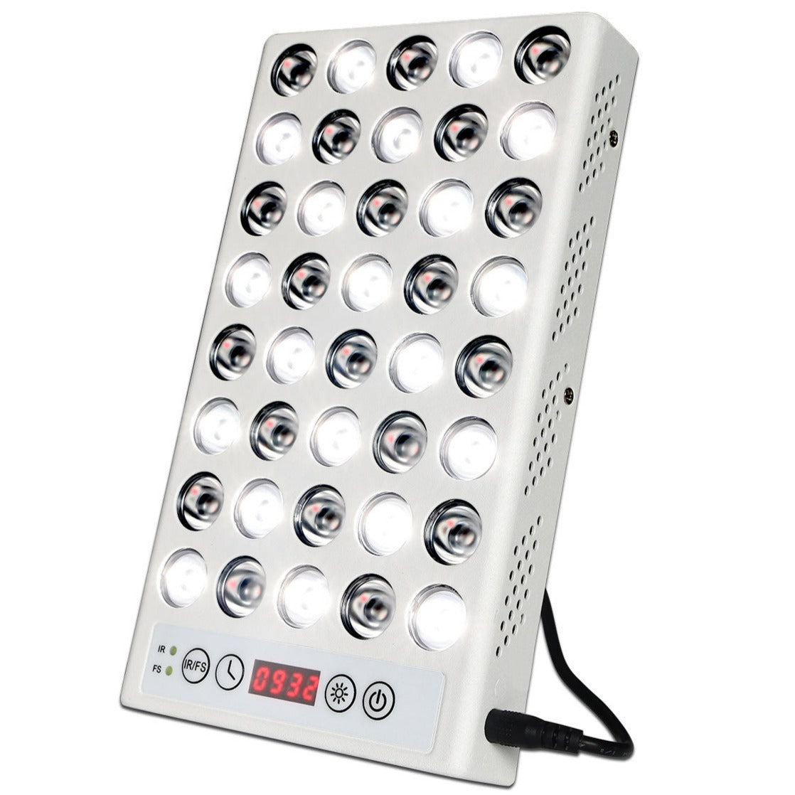
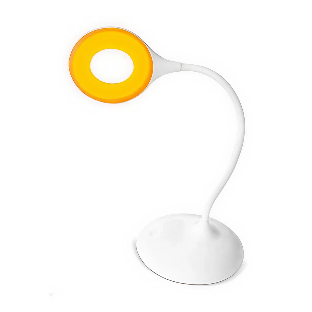
Red light therapy (PBM) at 660–850 nm restores ATP synthesis, improves blood flow, and reduces oxidative stress in demyelinated tissues. PBM enhances cytochrome c oxidase activity and supports remyelination in neurodegenerative diseases (Hamblin, 2016).
Grounding and cold thermogenesis restore zeta potential in cells, enhancing membrane potential and reducing inflammation. Cold exposure, particularly when combined with sunlight, induces mitochondrial biogenesis and promotes myelin repair (Lee et al., 2012).
Blood as Redox Proxy:
-
Poor RBC morphology and reduced deformability suggest intracellular redox collapse
-
Spectrin breakdown from UV deficiency reduces RBC pliability and reflects myelin stress
-
These changes are seen in MS and ALS patients before symptom onset (Visser et al., 2004)

Conclusion: Myelin Is Forged in Light
Demyelination diseases are not merely neurological, they are photonic. They arise when light is absent, redox is imbalanced, and mitochondrial coherence is lost. Mitochondria are the arc welders of biology. They forge life’s architecture through light, water, and magnetism.
The future of medicine lies not in synthetic repair but in reactivating our body's ancient blueprint for self-assembly, by returning to the natural environment it evolved in. With light as the first language of life, we reclaim not just our health, but our consciousness.
References
Campbell, G. R., Mahad, D. J. (2014). Mitochondrial dysfunction and axon degeneration in progressive multiple sclerosis. FEBS Letters, 588(22), 4606–4614.
Hamblin, M. R. (2016). Shining light on the head: Photobiomodulation for brain disorders. BBA Clinical, 6, 113–124.
Kruse, J. (2023). Decentralized Medicine Blog Series #47–52. [Online resource].
Lee, J., Duan, W., Mattson, M. P. (2012). Evidence That Brain-Derived Neurotrophic Factor Is Required for the Beneficial Effects of Exercise on Synaptic Plasticity and Cognitive Function. Frontiers in Aging Neuroscience, 4, 13.
Mattson, M. P., Liu, D. (2002). Energetics and oxidative stress in synaptic plasticity and neurodegenerative disorders. Neuromolecular Medicine, 2(2), 215–231.
Pierrot-Deseilligny, C., Souberbielle, J. C. (2017). Vitamin D and multiple sclerosis: An update. Multiple Sclerosis and Related Disorders, 14, 35–45.
Swerdlow, R. H., Burns, J. M., Khan, S. M. (2014). The Alzheimer’s disease mitochondrial cascade hypothesis. Journal of Alzheimer’s Disease, 20(Suppl 2), S265–S279.
Van Wijk, R., Van Wijk, E. P. A. (2005). Multi-site recording and spectral analysis of spontaneous photon emission from human body. Experientia, 61(5), 470–477.
Visser, E. J., Turturici, G., de Haan, A., et al. (2004). Blood rheology and RBC morphology in patients with ALS. Muscle & Nerve, 29(1), 110–113.
Zecca, L., Youdim, M. B., Riederer, P., Connor, J. R., Crichton, R. R. (2004). Iron, brain ageing and neurodegenerative disorders. Nature Reviews Neuroscience, 5(11), 863–873.


Disclaimer
The information on this site is provided by BioSpectral Systems for educational and informational purposes only. It is not intended to diagnose, treat, cure, or prevent any disease and has not been evaluated by the U.S. Food and Drug Administration or any other regulatory authority. Always consult a qualified healthcare professional before making any changes to your health regimen. By using this site, you acknowledge that you do so at your own discretion and agree that BioSpectral Systems, its affiliates, and contributors are not liable for any outcome resulting from the use of the information presented.
Share:
Folic Acid Trap: Fortified Food May Harm Brain Development
Why Clear Blue Light Gaming Glasses Aren’t Enough Protection